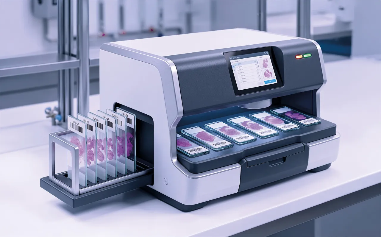
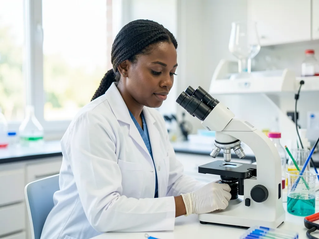
MyeloAID

MyeloAID — AI-assisted cell classification for bone marrow smears

MyeloAID automates the morphological differentiation of bone marrow smears, turning a manual, time-consuming process into a swift guided review. It produces a detailed, reproducible bone marrow differential in seconds and works with the hardware you already own.

Our edge

Built on the right expertise and the right data

Built by people like you

MyeloAID is designed by hematologists and MTAs, for hematologists and MTAs. We make sure the software fits how labs actually work instead of forcing a new routine.

Trained on the right data

Our models learn from curated, proprietary datasets of healthy and diseased bone marrow from leading German hematology centres, labelled by over ten morphology experts.

How it works

How MyeloAID fits your lab workflow

MyeloAID never runs on its own. It does the heavy counting, and a human expert always reviews and approves the result. It is also hardware-agnostic: upload images from the microscope camera or scanner you already have to the Cancilico cloud platform, and MyeloAID returns a structured bone marrow differential and a PDF report you can link back to your LIS. Pick the workflow that matches how your hematology service works today.

AI-generated illustration of a microscope fitted with a camera capturing fields of view Hybrid

Microscope camera

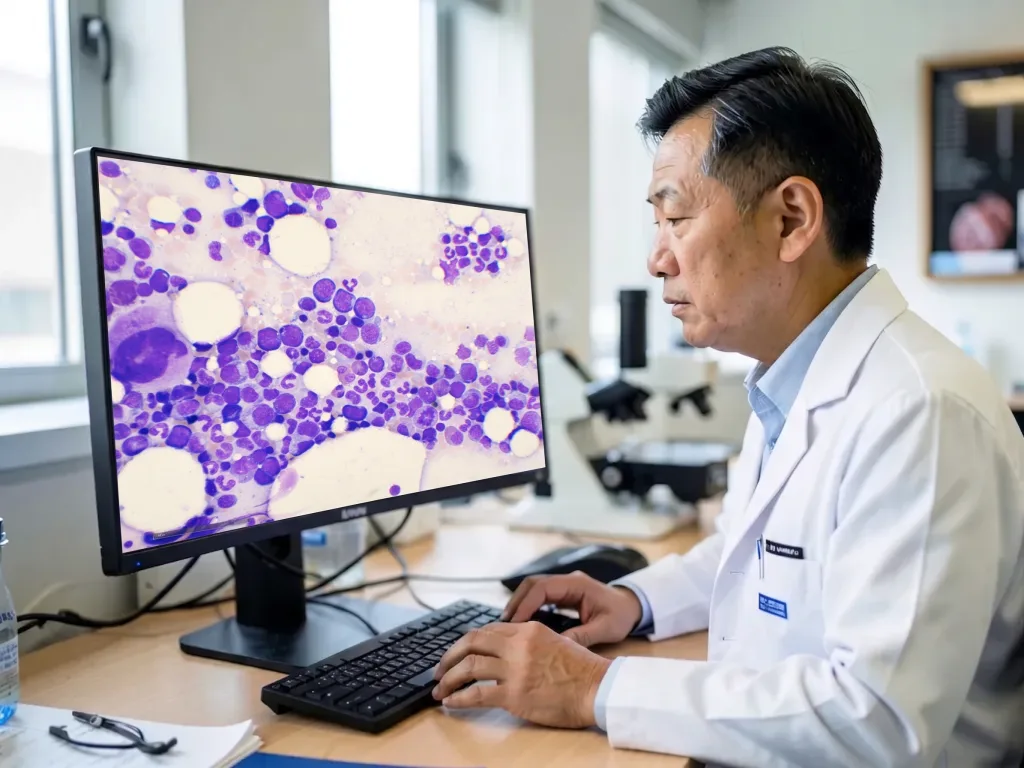
Keep working at the eyepiece. Your operator pans the slide and captures fields of view, and MyeloAID analyses the morphology as you go. It is a familiar way to work, with low up-front cost and a quick start. A good fit for low to medium volumes. No camera yet? We can advise and set you up with one for the best experience.

AI-generated illustration of a whole-slide scanner digitising slides in batches Fully digital

Whole-slide scanner

Scan slides in batches and let MyeloAID run across the whole slide. It automatically selects regions of interest for you to accept or override. The results are reproducible, it scales to large teams, and it supports remote review. A good fit for high-volume hospitals and reference labs.

Either way, you get the same structured bone marrow differential and PDF report. We validate MyeloAID against common scanners and image formats, so for supported hardware new cases can land in the platform automatically. Tell us your scanner model and case volume, and we’ll tell you what to expect.

AI-generated portrait of a laboratory technician reviewing a bone marrow smear at a camera-equipped microscope

For lab technicians

MyeloAID does not replace you. It takes the bulk of the repetitive counting off your hands, so you can focus on the difficult cases that need a trained eye. You stay in the driver’s seat: review what the AI proposes, and change the analysed regions or cell classes whenever your expert judgement says otherwise.

AI-generated portrait of a hematologist reviewing digital microscopy slides on screen

For hematologists & hematopathologists

Manual differentials are slow, and two readers, or the same reader on two different days, can score the same slide differently. MyeloAID applies the same criteria every time, so it reduces inter- and intra-reader variability and gives you reproducible cyto-morphological analysis. You still own the diagnosis; the AI accelerates the counting so you can spend your time on interpretation and treatment decisions.

AI-generated portrait of a laboratory director in a modern hematology lab

For lab directors

Because MyeloAID works with the imaging hardware you already own, you can add AI-assisted morphology without buying a six-figure scanner. It helps you control costs and take on more cases as demand grows, while keeping your reporting consistent across the lab.

Regulatory status

MyeloAID is currently available for research use only (RUO); not for use in diagnostic procedures. We are working toward IVDR and FDA clearance.

Bring AI-assisted morphology to your lab

Tell us your scanner model and case volume, and we’ll show you what MyeloAID can do on your own slides.